
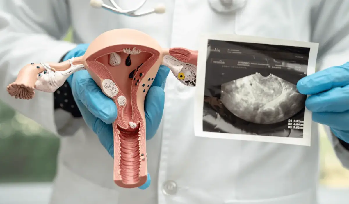
Cervical cancer is a highly preventable condition because of the highly reliable screening methods available for it. Although not typically used, ultrasound can also be used to detect cervical cancer sometimes.
But how reliable is ultrasound for screening cervical cancer? How much information can be collected through an ultrasound of cervical cancer? Compared to other methods, how accurate is an ultrasound for cervical cancer screening?
If a doctor finds something unusual in your symptoms or test results, they might recommend an imaging study to make an accurate diagnosis. However, ultrasound is not usually chosen by doctors.
What Is an Ultrasound: Can It Detect Cervical Cancer?
An ultrasound or sonogram uses high-frequency sound waves to capture real-time images and videos of the inside of your body. During an ultrasound, your healthcare provider will apply a gentle, water-based gel to your skin and then glide a handheld device, called a transducer or probe, that sends inaudible, high-frequency sound waves into your body. The sound waves bounce back after reaching your organs and tissues as echoes. A computer translates these echoes into images on the screen.
An ultrasound is one of the best imaging tests that helps to look at what is happening inside the body. In obstetrics and gynecology, it is commonly used to monitor fetal development. It also helps to identify fibroids, cysts, and other growths. However, for the screening of cervical cancer, ultrasound is not an ideal method.
Since there are more advanced screening and diagnostic methods for cervical cancer and ultrasound is not generally used as a cervical cancer screening method, it is unlikely for a doctor to identify cervical cancer while imaging for other purposes. Ultrasound technicians are not usually trained to recognize cervical cancer.
However, an ultrasound might be used in the later part of the diagnosis of cervical cancer as a supporting method, since several imaging tests might be required to check if the cancer has spread to nearby tissues or organs. For that, your doctor might use a transvaginal ultrasound and a transabdominal ultrasound.
In a transvaginal ultrasound, a probe is inserted into the vagina to get a clear and detailed view of the cervix, helping to detect if the cancer has spread to the tissue. Whereas in a transabdominal ultrasound, the probe is used to glide over the abdomen, giving a view of the broader pelvic area.
An ultrasound may sometimes be used to investigate a suspected mass. It can be used to reveal a hypoechoic (darker) or heterogeneous mass in the cervix, increased blood flow in the tumor (using a color Doppler ultrasound), the exact size of the tumor, and whether it has invaded the vagina or adjacent pelvic organs. But it can never be used to diagnose cervical cancer.
What Tests Are Required to Diagnose Cervical Cancer?
The proper diagnosis of cervical cancer is done through a few screening tests and diagnostic tests. The screening tests for cervical cancer include:
Pap Smear: A Pap smear, or a Pap test, helps detect abnormal changes in the cervical cells. In this test, the healthcare provider will collect a few cells and mucus from the cervix using a swab or a spatula and examine the sample in a laboratory.
A Pap smear, which is part of the routine physical exam, is the best preventive method for cervical cancer since it can detect abnormalities even at the precancerous stage, allowing early diagnosis and treatment. A complete recovery or prevention of cervical cancer is possible in most cases that are identified through the routine Pap smear.
HPV Test: An HPV test identifies the presence of human papillomavirus, which is the most common cause of cervical cancer across the world. This test is also part of your routine physical exam. There are over 200 types (strains) of HPV that cause infection in humans, and most of them are asymptomatic. Although the low-risk HPV strains only cause genital warts, persistent infection of high-risk strains can cause abnormal changes in the cells.
Colposcopy: If the doctor finds abnormalities in the Pap smear and HPV test, they will recommend a colposcopy for a detailed analysis. This is not much different from the physical examination. The doctor will insert a spatula into your vagina to have a closer and more detailed view of your cervix.
The Diagnostic Tests for Cervical Cancer
For the confirmation of cervical cancer, a biopsy is required. Your doctor will recommend a biopsy if the colposcopy results are abnormal. This involves the detailed analysis of a small cervical tissue sample in a laboratory.
Colposcopy Biopsy: Sometimes doctors conduct a biopsy during colposcopy. They will perform colposcopy, take a small tissue sample if they identify an abnormal area in the cervix, and send it to the lab for detailed investigation.
Cone Biopsy (Conization): During this, the doctor will remove a cone-shaped section of your cervical tissue and send it to the lab. In the case of early-stage cervical cancer, this is sometimes performed as a treatment method.
Endocervical Scraping (Endocervical Curettage): This involves removing tissues from the endocervical canal. It is performed as a follow-up test when the results from other biopsies are inconclusive.
Conclusion
An ultrasound scan is a very good imaging test to view the inside of one’s body. Ultrasound can also sometimes detect cervical cancer. However, it cannot replace the regular screening and diagnostic process for cervical cancer.
For the accurate diagnosis of cervical cancer, the detailed examination of the cervical tissues is very crucial. Many advanced screening and diagnostic tests can help confirm cervical cancer more precisely compared to an ultrasound. An ultrasound is only helpful in the later stages of diagnosis and treatment, as a supporting imaging method.
FAQ
Yes. Cervical cancer can be detected through an ultrasound. A transvaginal ultrasound can sometimes help visualize large tumors or abnormal tissue growth in the cervix. It can also help measure the tumor size and depth of its invasion.
Although advanced cervical cancer can be detected through various symptoms, early-stage cervical cancer rarely shows symptoms. However, routine screenings like a Pap smear or an HPV test help early detection of cervical cancer or its possibility.
During your pelvic exam, your doctor will gently insert a spatula into your vagina and open it to expand your vaginal walls. Using a brush, swab, or a spatula, a few cervical cells will be collected and tested in the lab to detect abnormal changes.
Stage 1 cervical cancer is the primary stage of cervical cancer, where the tumor is located only in the cervix and has not spread to the nearby pelvic tissues, lymph nodes, upper vagina, or distant organs. This stage is highly curable.